 You may have noticed that I didn't mention the study in my last post about my doctor visits. That's because it deserves a post all its own.
I was told that I would be participating in a study about eye movement and tracking, and that because I had lost my vestibular nerve on one side, it would be helpful to see how my eyes and brain had adapted so far. I would have to wear contact lenses with sensors around the outside of them that would monitor my eye movement while my head turned from side to side. I said, "OK, sounds doable."
When we walked into the lab with that crazy contraption, I thought, "What in the world did I sign up for???"
Well, I signed up to particpate in a study called "VOR adaptation and the use of saccades as rehabilitation strategies;" a quick Google search reveals that VOR stands for vestibulocular reflex and that saccades are fast movements of the eye.
The consent form explains that "this research is being done to better understand how the vestibular part of the inner ear plays a part in vision and balance. The vestibular part of the inner ear senses your head tilt and rotation during movements like walking or driving. It sends information to the reflexes that help keep your eyes looking straight ahead while you are moving. When the system fails, abnormal reflexes can cause dizziness and blurred vision."
Yep, that's what's been happening to me since the surgery - quick movements of my head make me a little unsteady and cause my vision to bounce around a bit. Sounds like it's going to be hard to do well on these tests...
After everything was thoroughly explained to me by the lead researcher and I signed the consent forms, we got started. I sat in the chair, which Phil and I later found out was built in 1960 and is one of about ten in the world, and proceeded to have contact lenses positioned on my eyeballs. They weren't for vision though - the center was open, and a very fine wire around the perimeter of the lens connected to a recording system that measured magnetic fields in the room.
Additionally, I was fitted with a bite block made of dental putty so that my head movement could be measured with another sensor inside the block. If you refer to the photo above, you will see a large metal frame surrounding the chair. This is where the magnetic field originates from, and the most concentrated point of magnetic activity is focused around the head area (it was a very weak magnetic field and did not require me to remove my insulin pump).
We began the Dynamic Visual Acuity testing. This consisted of me sitting in the chair with the lenses and bite block in place, looking at a computer screen several feet in front of me. First we did the test while my head was stationary; what I needed to do was identify the way a letter E was facing (up, down, left, right) at a variety of sizes from large (~2 inches) to small (~1/2 inch). This was not difficult when I wasn't moving.
The lead researcher stood behind me, placed another sensor band around my head, and then started the motion part of the test. He quickly moved my head with his hands, which activated the letter E to appear on the screen. My job was to again identify the way it was facing - the problem was that unless the E was at its largest size, it just looked like a blur to me. This is because the vestibular function of my left ear has been disabled, and my brain has not been fully trained to compensate yet. What happens is that when I look at a fixed object and my head turns rapidly, my eyes move along with my head for a split second until they re-fixate on the object. This is not how it is supposed to work; the eyes would remain fixated on the object no matter how fast the head is moving.
From what I could tell, I was better able to identify the position of the E when my head was turned to the right, which makes sense since my right-side vestibular function is intact. We repeated this test several times on each side to account for each direction that corresponds to a different semicircular canal in the eardrum. There was straight left-to-right, straight up-and-down, diagonal left-to-right, and the others for the right side as well. I asked Phil afterward if it was obvious which way the E was facing when I wasn't able to tell, and he said yes.
After the lead researcher reviews the results, he is going to send them to me so I can see where my deficiencies are. I have a feeling that I'm definitely going to want to see a vestibular therapist...I doubt ping pong is going to be a miracle cure.
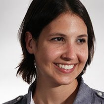
 And, yes, it was awkward wearing the wired lenses. The right one got out of position halfway through and he took it out, so my left eye was doing all the work. Fortunately, I did not get a corneal abrasion from the testing, which was one of the risks, nor did I suffer a neck injury from all of the twisting. The photo at the left was taken while my eyes were being checked for scratches with fluorescent drops and a black light. If you look closely, you can see my eyes fluorescing...interesting, huh?
All in all, I think this was a worthwhile thing to do, and although they say in the consent form that there is no direct benefit to the participant, I think there just may be for me.

No comments:
Post a Comment